Membrane Proteins
Biophysicist Georg Büldt on X-ray crystallography, structure of proteins, and free electron laser
videos | August 23, 2016
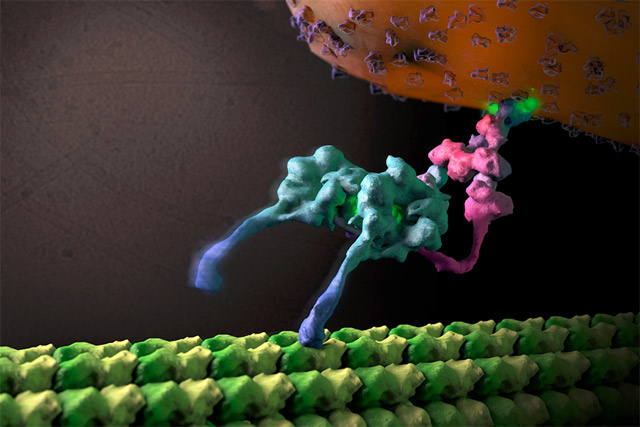
Since we know that all living matter consists of cells, of course, membrane proteins, or membranes at all are in the focus. And of course, the cell must have communication from the outside to the inside and must also transport materials from the inside to the outside. And this goes over the membrane which normally protects the cell from things from the outside, and also that the inside doesn’t go from itself to the outside. And all this is done by membrane proteins, therefore membrane proteins are very important.
Unfortunately, it took a very long time until one was able to study membrane proteins and I can tell you that normally, when you want to study a protein, the basis is that you know it’s structure. Structure of a protein you see by the x-ray crystallography normally, in former times at least. And it took a long time until the membrane protein structure came out. The first membrane protein that people were working on comes from the archaebacteria that was Bacteriorodopsina.
And people around the world tried for forty years to crystallize it, and it wasn’t possible until ten years ago or fifteen years ago. And then it starts that many membrane proteins could be crystallized because a certain method was discovered.
And since that time we know a lot more about membrane proteins, although it is still only compared to water soluble proteins, only five percent of that.
Although these proteins are so important, because to see how communication works over the cell is very important, and also the transport of materials. Just to tell you, that from the first membrane proteins which were solved that was involved in drug discovery. These proteins can be easily manipulated by drugs from the outside: you just put in the blood, or you eat something of this drug, and it binds to the signaling protein and then it stops to do this. Or it increases its business. And this, as you see, is important for medicine and for pharmaceutical companies. Therefore, in the United States, where they really started this commercialization of it, billions of dollars were coming from such research in companies which take these things up.
I said already, that the structure of a protein is the basis on which you can work but to really find out how it works, you need a lot of methods. And one important method is, of course, infrared spectroscopy. Because, for instance, in many cases you can follow up in what part of the protein conformation changes occur. This is important because a protein when it is folded – normally protein is a linear stretch of amino acids – and it becomes a machine when it is folded. And to understand this machine you often need spectroscopic methods. When you know the basis by a structure then you can follow up by the spectroscopic methods where in this structure – say, you have a protein like rhodopsin here in the eye, you give a flash of light, and then you look what happens in time when this flash of light comes to the rhodopsin in the eye which gives you the vision, and what occurs in time. Then this spectroscopic method can really tell you what happens in what part of the protein, and then you know the function.
Of course, this is also important for drug discovery in other cases. You can do it in many cases, but it’s not only that, you can also do NMR.
What is the advantage of NMR to x-rays? In NMR you can study proteins which are not in the crystal. When you have a protein in the crystal the conformation changes outside the protein are hindered off by the packing. And therefore in NMR experiment you have it all in solution, so the protein is free to do its conformation changes.
On the other hand, in the NMR experiment of a bigger protein, say, forty-kilodalton takes two years or three years, whereas to find a structure when you have crystals, is one week. So you see already the difference. But one has to say that x-ray crystallography was invented in Germany in the beginning of, say, 1900 by Laue.
And then Bragg in England continues on working on natural substances, and now we are much ahead. Maybe in 50 years NMR is very strong and then many people will go to the NMR. But now another method is coming up that is free electron lasers.
The X-ray crystallography has one bad point, and that is radiation damage. When you put an X-ray beam on a crystal, after a while it is gone. So people were thinking:”Сan we prevent from this?” And so they invented a free electron laser.
That is something which can make x-ray pulses, which have length of 1 femtosecond to 3 hundred femtosecond. On your wish: say you want 10 femtosecond then they can make it 10 femtosecond. And they put in this pulses a lot of photons, of x-ray photons. And they go then within, say you make a pulse of 10 femtosecond, they only evaluate your crystal for 10 femtosecond. And then it is destroyed because the number of photons is very high, but the reflexions, the information is already on the detector before it falls in peaces. So you see, that is the way we are going because the thing is that, maybe in far future, we need not to have any crystals, and we can do it on a single protein. People dream of it, that then they will have more and more… But of course there is a limit. You can not press electron bunches which are in this free electron laser together. The free electron laser gives photons which are coming from electron bunches which are in the magnetic field always changing the direction. And when they change the direction, photons are emitted. And this is the principle.
And it would be of course interesting to see one day which method gives most of the impact, most of the information. And so far I say it’s really x-ray crystallography that gives most of the information but in future it may change, and NMR can come up. And I don’t know whether really the dream of free electron laser will be fulfilled in future, then of course, that will give us a lot of information because you have a single protein then in the beam. And I think in future, for instance there is one sort of membrane proteins like G protein-coupled receptors which are very important for giving a signal to the nuclears in the cell.
And these G protein-coupled receptors, we have 8 hundred of them. But we know now only from 20 of them the structure. We will never solve this 8 hundred structures but they are in groups, and we would like to have from each group a structure, then we can model the other structures.
And that is the way we like to proceed and we are working in this way together with Scirpps in California. And Scirpps is also in collaboration with Shanghai in China, because also the Chinese people see that there is a lot of impact of this thing in terms of medical treatments and drugs discovery and all this things.


