Short-Term Memory
Neuroscientist Neil Burgess on the difference between short-term and long-term memory, phonological loop, and ...

On June 19, 2015, the eLife journal published a paper titled “The mesh is a network of microtubule connectors that stabilizes individual kinetochore fibers of the mitotic spindle”, describing the network of microtubule connectors which gives structural support to dividing cells. We have asked one of the authors of this research, Assoc. Prof. Stephen J. Royle from the University of Warwick, to comment on this work.
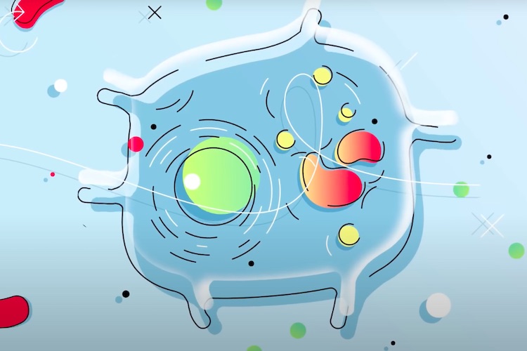
When a cell divides, the two new cells need to get the right number of chromosomes. If this process goes wrong, it is a disaster which may lead to disease, e.g. cancer. The cell shares the chromosomes using a “mitotic spindle”. This is a tiny machine made of microtubules and other proteins. We have found that the microtubules are held together by something called “the mesh”. This is a web-like structure which connects the microtubules and gives them structural support.

Faye Nixon, a PhD student in the lab did most of the work. She used a method to look at mitotic spindles in 3D to study the mesh. My lab actually discovered the mesh by accident. A previous student, Dan Booth – back in 2011 – was looking at mitotic spindles to try and get 3D electron microscopy (tomography) working in the lab. Tomography works just like a CAT scan in a hospital, but on a much smaller scale.
The mesh is found in the gaps between microtubules that are 25 nanometre wide (1 nanometre is 1 billionth of a metre). This is about 3,000 times smaller than a human hair. It was Dan who found the mesh and gave it the name. Other people in the lab did some really nice work which helped us to understand how the mesh works in dividing cells. Cristina Gutiérrez-Caballero did some experiments using a different type of microscope and Fiona Hood contributed some test tube experiments. Ian Prior at University of Liverpool, co-supervises Faye and helped with electron microscopy.
All cell biologists dream of finding a new structure in cells. It’s so unlikely though. Scientists have been looking at cells since the 17th Century and so the chances of seeing something that no-one has seen before are very small. In the 1970s, “inter-microtubule bridges” in the mitotic spindle were described using 2D electron microscopy. What we have done is to look at these structures in 3D for the first time and find that they are a network rather than individual connectors.
Some human cancer cells have high levels of proteins called TACC3 and Aurora A kinase. We know that TACC3 is changed by Aurora A kinase. This changed form of TACC3 is part of the mesh. In our paper we mimic the cancer condition by increasing TACC3 levels. The mesh changes and the microtubules become wonky. This causes problems for dividing cells. It might be possible to target TACC3 using drugs to treat certain types of cancer, but this is a long way in the future.
We need to identify more of the proteins that make up the mesh. We have identified one group of proteins (including TACC3) but there are others in there. There have been clues from labs around the world for potential mesh proteins. We are testing if any of these are mesh components and are developing ways to distinguish these proteins in the electron microscope.

Neuroscientist Neil Burgess on the difference between short-term and long-term memory, phonological loop, and ...

Neuroeconomist Sacha Bourgeois-Gironde on the symbolic aspect of money, brain processing of economic artifacts...

University of Strasbourg professor, Burkhard Bechinger, on discovering new antibiotics, the interaction of pep...