Microrobotics
Computer Scientist Sergej Fatikow on the variety of microrobots, possibilities for their application, and curr...

Computed Tomography (CT), often linked with medical research and diagnostics, is not limited to these fields. It is a non-destructive method that opens up new possibilities for quality control, research, and development in various industries. With CT, one can study the internal structure of objects without causing any physical damage, detect hidden defects, and analyze materials with a high degree of detail.
Tomography is a method that allows us to look inside an object without destroying it. To achieve this, the object is illuminated using X-ray radiation, and a series of images are taken from various projection angles. Images are formed as the radiation passes through the object—the attenuated X-rays fall on recording devices, creating a picture.
X-ray radiation can restore several images, each taken from a different angle. A mathematical reconstructor can restore the surface and the scanned object’s internal morphological structure. The resulting image is a three-dimensional digital representation of the examined item. X-ray laboratories usually use a spectrum of wavelengths instead of a single wavelength to speed up the image acquisition process.
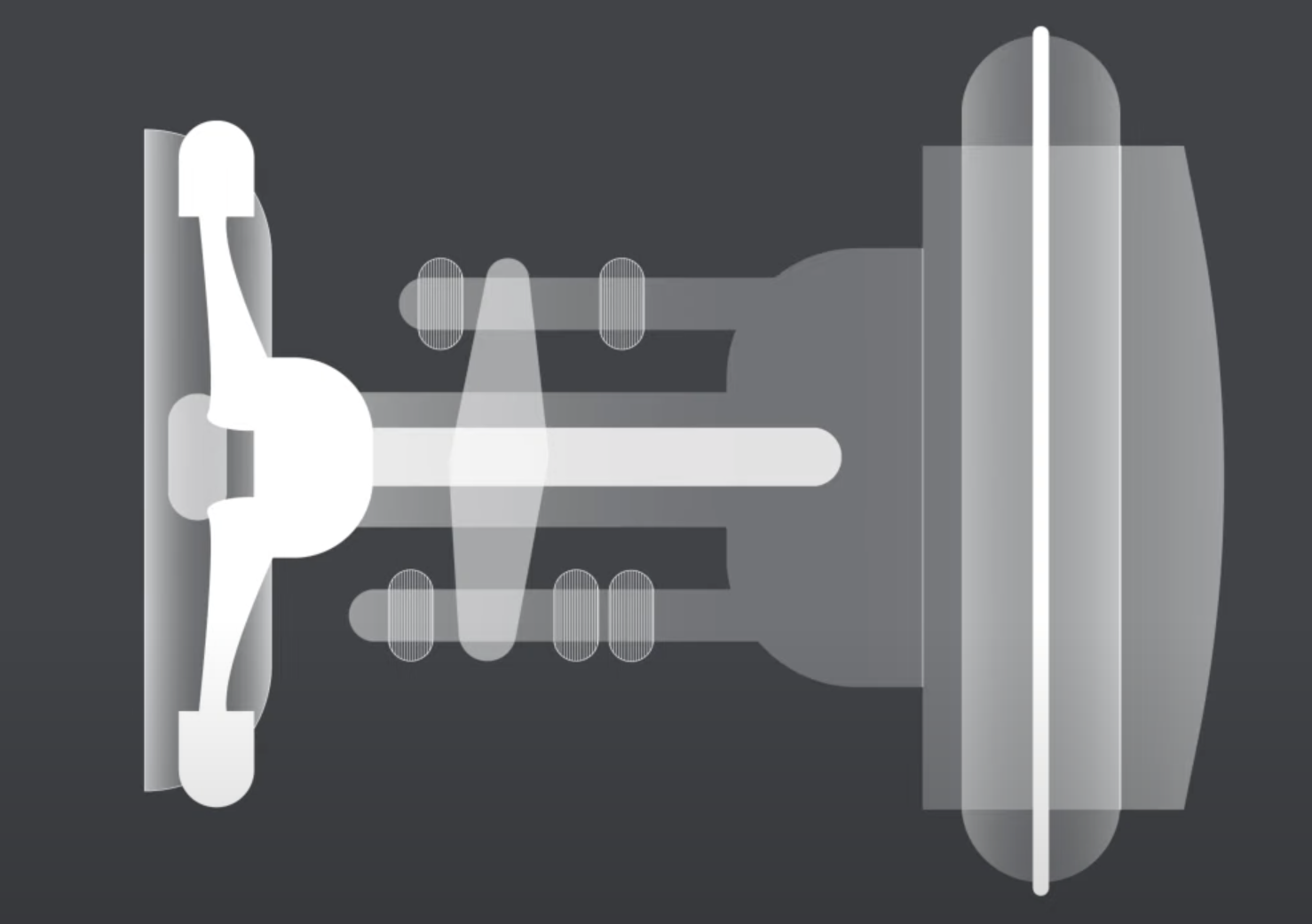
A basic tomograph consists of three elements: a source, a receiver, and a schematic. If the object under study is small, it can be fixed on a rotating platform. In this case, the “source-receiver” pair remains static. Alternatively, the sample remains stationary on a unique stand while the “source-receiver” pair rotates around it. This method is often used in robotic tomographic devices.

Although tomography is primarily known in medicine, it is one of the main tools for examining the internal structure of products in industry.
For example, when constructing a complex product consisting of many parts, each can be placed in a tomograph to ensure they are in perfect condition. This allows for quality control before the parts are assembled. The exterior might look perfect once the parts are joined using materials like plastic and ceramics, but internal structures can develop cracks. Subjecting such products to pressure or temperature stress is not feasible. With computed tomography, instead of disassembling and reassembling the product, you can scan it immediately to identify the problem. This enables the replacement or repair of the defective part without needing dismantling.
Tomography is used in various industries, from heavy industry to microelectronics, and even for developing new technological processes. It provides a non-destructive way to study the internal structure of objects, ensuring their integrity and functionality without compromising their form.
To see and interpret the internal structure of objects, a “brain” is needed to make decisions. This role is taken on by artificial intelligence (AI)—software that performs several vital tasks in tomography. AI organizes the assembly of images, which the reconstructor will later process. It handles the reconstruction, which involves restoring a digital three-dimensional structure from a set of images called radiograms. This task is the most complex because the data is often insufficient.
The third function of AI is the automatic analysis of the obtained image. AI compares it with an existing digital copy of the object created from blueprints or with a ready-made model of the product if it was made using additive technologies. In this way, AI determines where and what problem has arisen: whether a crack has appeared, the object has changed dimensions under pressure, or an electromagnetic field has caused a change in the physical structure, etc.
Subsequently, AI automatically generates a conclusion and a report on the study conducted. Thus, the software acts as the “brain” of the tomograph. By integrating AI, computed tomography becomes more efficient and accurate, enhancing its capabilities in medical and industrial applications.
The primary limitation of computed tomography (CT) is the need to illuminate the object without damaging it. When dealing with materials like steel, for example, in spacecraft construction, the amount of X-ray radiation is not an issue because such materials are not sensitive to radiation. However, some devices can be damaged by X-ray exposure. This is particularly true for small objects studied at high resolution, which can easily be spoiled because X-rays can dislodge electrons. If the object is a microstructure that operates on these very electrons, it cannot be damaged.
This limitation is known as the dose load limitation. The amount of projection data collected becomes crucial: On one hand, it is necessary not to damage the object, and on the other, it is essential to gather enough data to reconstruct its internal digital analogue with the required accuracy. For instance, more than two images taken at 0 and 90 degrees are usually required for an accurate internal reconstruction of the entire object.
Traditionally, imaging protocols dictated when measurements began and ended, were analyzed, and transmitted for reconstruction. These protocols did not account for scenarios where the object moved or began to melt, as in the case of ice objects, making it impossible to compile the image accurately. Monitoring reconstruction helps detect and address this problem.
One of the critical advancements in computed tomography (CT) is reducing the radiation point size, which allows for higher resolution. This means that the voxel size in the digital image will be smaller. Ideally, having a point radiation source would be perfect, but creating a source that emits many X-ray quanta from a small point is very challenging. Measurements must be conducted quickly without taking too much time, so researchers are working on developing powerful yet compact X-ray sources.
Additionally, different research tasks require various ranges of X-ray radiation, from a few keV to several hundred keV. For example, alloys cannot be penetrated with an energy of 1 keV; hundreds of kiloelectronvolts are needed to penetrate metal. Conversely, only a few keV are sufficient for plastic, as the likelihood of interaction with higher energy radiation is extremely low. This diversity in research needs underscores the necessity to develop sources capable of providing a wide spectrum of radiation, highlighting the versatility and adaptability of CT technology.
Describe advancements in high-resolution imaging and analysis, and consider a situation where an object like a mobile phone needs to be examined with extremely high detail. The phone’s size might be approximately 15 by 10 centimetres, and in some cases, smaller, such as 10 by 7 centimetres. The main goal is not just to observe the smallest components of the device, such as microchips, but to do so with nanometer resolution, showcasing the precision and detail CT technology can provide.
Many are eager to observe how dynamically mobile objects change over time. A significant interest has emerged in 4D tomography: numerous scientists and researchers claim they are already applying this technology. By 4D tomography, we mean not just visualizing an object in three dimensions but adding time as the fourth dimension.
A complete tomographic scan can be conducted multiple times if an object changes so slowly. Then, the collected data is compiled into a sequence, creating a video; this is 4D tomography. In this case, there is a unique opportunity to study an object at a single moment and observe its evolution.
The ultimate goal is to develop algorithms that allow changes in the object to be captured in a single tomographic scanning cycle. This means using a limited number of projections. Thus, several projections enable capturing one phase of change, and subsequent projections capture the next phase. As a result, a “movie” of the object’s transformations throughout one scan is obtained.
This approach has significant advantages, particularly in reducing the dose load on the object. Instead of repeatedly exposing it to X-rays, one measurement cycle is used to obtain dynamic information. This means that our algorithm extracts the maximum useful information from a minimal number of projections.

Computer Scientist Sergej Fatikow on the variety of microrobots, possibilities for their application, and curr...

Biographer and historian of science Naomi Pasachoff on the discovery of radium and polonium, acquaintance with...

Prof. Emery Brown of MIT on the rapid eye movement stage of sleep, electroencephalogram patterns, and the role...