


In the 19th century, invisible radiation capable of passing through flesh and other materials seemed utterly unbelievable. Now, X-rays are widely used for creating medical images, conducting radiation therapy, analyzing works of art, and solving problems in atomic energy. Together with physicist Alexander Dolgov, we explored how X-ray radiation was discovered and how it helps people.
Röntgen’s discovery

One of the scientists who worked passionately during this period was Professor of Physics and Rector of the University of Würzburg, Wilhelm Conrad Röntgen. On November 8, 1895, he stayed late in the laboratory, as often happened, and decided to conduct experimental research on electrical discharge in glass vacuum tubes. He darkened the room and wrapped one of the tubes in opaque black paper to make observing the optical phenomena accompanying the discharge easier. To his surprise, Röntgen saw a fluorescence strip on the nearby screen, covered with crystals of barium platinocyanide.
The scientist hardly could have imagined then that he was on the threshold of one of the most important scientific discoveries of his time. Over a thousand publications were written about X-rays the following year, and medics immediately adopted the invention. This later led to the discovery of radioactivity and new scientific disciplines.
Röntgen dedicated the following weeks to investigating the nature of the mysterious glow and established that fluorescence appeared every time he applied current to the tube. The radiation source was the tube rather than any other electrical circuit part. Not knowing what he had encountered, Röntgen decided to refer to this phenomenon as X-rays.
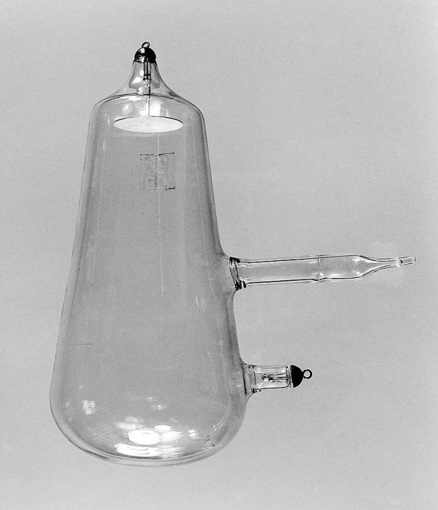
Crookes tube — a device that was used to produce X-rays for the first time unintentionally // wikipedia.org
Röntgen later discovered that this radiation could penetrate almost all objects to varying depths, depending on the object’s thickness and the material’s density. For example, a small lead disk between the discharge tube and the screen proved impenetrable to X-rays, while the bones of the hand cast a darker shadow on the screen, surrounded by a lighter shadow from soft tissues. Soon, Röntgen discovered that X-rays caused the screen covered with barium platinocyanide to fluoresce and darkened photographic plates (after development) in areas where X-rays hit the photographic emulsion.
Röntgen confirmed that he discovered radiation previously unknown to science during his experiments. On December 28, 1895, he reported the results of his research in the article titled “On a New Kind of Radiation” in the journal “Annals of Physics and Chemistry.” At the same time, he sent famous photographs of his wife Anna Bertha Ludwig’s hand to scientists. Thanks to Röntgen’s old friend, Austrian physicist Franz Exner, these photos were first seen by the residents of Vienna on January 5, 1896, in the newspaper Die Presse. The next day, news of the discovery reached the London Chronicle. Thus, Röntgen’s discovery gradually began to enter people’s everyday lives. On January 20, 1896, in New Hampshire, doctors assisted a person with a broken arm for the first time with a new diagnostic method — an X-ray.
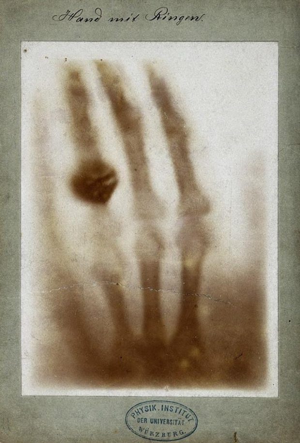
X-ray image of Anna Bertha Ludwig’s hand // wikipedia.org
Early use of X-rays
By 1900, just five years after its discovery, the use of X-rays in diagnostics was considered an integral part of medical practice. The statistics collected by the oldest hospital in Pennsylvania are a significant indicator of the dissemination of X-ray radiation technologies. According to the study, in 1900, only about 1–2% of patients received assistance using X-rays. By 1925, this number had risen to 25%.
At that time, X-rays were often used in unusual ways. For example, to remove hair. People considered this method to be less painful than tweezing or waxing. Also, X-ray radiation was used in pedoscopes —devices for fitting shoes. These were X-ray machines with a special recess for the feet and windows through which the customer and the salespeople could assess how the shoes fit.
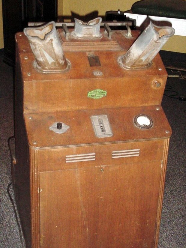
Fluoroscope for shoes // wikipedia.org
The nature of X-rays
X-rays can be classified into soft and hard radiation, with a conventional boundary on the wavelength scale at approximately 0.2 nm, corresponding to a photon energy of around 6 keV. Due to their short wavelength, X-rays are both penetrating and ionizing, as they interact with electrons when passing through matter, knocking them out of atoms. This interaction breaks the atoms into ions and electrons, altering the material’s structure.
X-ray radiation causes the luminescence of chemical compounds, known as fluorescence. When atoms in a sample are irradiated with high-energy photons, electrons are emitted and leave the atom. This creates “holes” or vacancies in one or more electron orbitals, causing the atoms to enter an excited and unstable state. Within millionths of a second, the atoms return to a stable state as vacancies in the inner orbitals are filled by electrons from the outer orbitals. This transition is accompanied by energy emission in the form of secondary photons, which is the fluorescence source.
X-ray Astronomy

One of the first X-ray sources in the sky, Cygnus X-1, was discovered in 1964, and today, most scientists are convinced that it is a black hole with a mass of about 15 solar masses // NASA
These cosmic X-ray radiation sources do not constitute a noticeable part of the natural radiation background and pose no threat to humans. The only potential exception would be a source of hard electromagnetic radiation, such as a supernova explosion occurring sufficiently close to the Solar System.
How to Create X-rays Artificially

X-Ray Tube // Serious Science
The continuous or pulsed X-ray tube remains the most common X-ray radiation source, but it is not the only one. High-intensity radiation pulses are generated using high-current discharges, where the plasma channel of the current is compressed by its own magnetic field—a process known as pinching. If the discharge occurs in a medium of light elements, such as hydrogen, it acts as an effective electron accelerator due to the electric field generated within the discharge. This field can significantly exceed the one created by an external current source. This method produces pulses of hard X-ray radiation with high-energy photons (hundreds of kilo electronvolts) with high penetrating power.
Electron accelerators, such as synchrotrons, produce X-ray radiation across a broad spectral range. In synchrotrons, radiation is generated inside a ring-shaped vacuum chamber, where a tightly focused beam of high-energy electrons moves along a circular orbit, accelerated to nearly the speed of light. As the electrons turn under the influence of a magnetic field, they emit photon beams tangentially to the orbit. These beams span a wide spectrum, with a peak in the X-ray range.
How to Detect X-rays
Nowadays, electronic detectors are most commonly used to register X-ray radiation. These devices produce an electrical pulse when a radiation quantum is absorbed within the detector’s sensitive volume. They differ in how they convert the absorbed radiation energy into electrical signals. X-ray detectors with electronic registration can be divided into ionization detectors, which operate based on the ionization of matter, and radioluminescent detectors, including scintillation detectors, which use the luminescence of matter under ionizing radiation. Depending on the detecting medium, ionization detectors are further classified into gas-filled and semiconductor detectors.
The main types of gas-filled detectors are ionization chambers, Geiger counters (Geiger-Müller counters), and proportional gas discharge counters. Radiation quanta entering the detector’s working medium cause gas ionization and the flow of current, which is then registered. In a semiconductor detector, electron-hole pairs are formed under the influence of radiation quanta, allowing an electric current to flow through the detector body.
A secondary electron multiplier is an open vacuum device (operates only in vacuum conditions) where incoming X-ray radiation is converted into a stream of primary electrons and amplified through secondary electron emission as they propagate through the multiplier channel. Microchannel plates work on the same principle. These plates consist of many individual microscopic channels that penetrate the plate detector. They can also provide spatial resolution and form an optical image of the cross-section of the X-ray beam hitting the detector by bombarding a phosphor-coated semi-transparent screen with the outgoing electron stream.
X-ray Radiation in Medicine
A relatively new field that is currently developing in microbiology and medicine is the use of soft X-ray radiation. When penetrating a living organism, it allows for imaging blood vessels, detailed examination of the structure of soft tissues, and even conducting microbiological research at the cellular level. An X-ray microscope that uses radiation from a pinch discharge in heavy element plasma enables the observation of details in the structure of living cells that even an electron microscope cannot see, even in specially prepared cellular structures.
One type of radiation therapy used to treat malignant tumours utilizes hard X-ray radiation. This is possible due to its ionizing effect, which destroys the tissue of the biological target. In this case, an electron accelerator is used as the radiation source.
X-ray Imaging in Engineering
The ability to penetrate materials makes possible X-ray imaging. This visualization method allows for displaying the internal structure of opaque objects, such as those made of metal. It is impossible to visually determine whether the welds on a bridge are secure, if the seam of a gas pipeline is airtight, or if the rails fit closely together. Therefore, in the industry, X-ray imaging is used for defectoscopy, which inspects the reliability of an object’s primary working properties and parameters or its individual elements without taking the object out of operation or dismantling it.
X-ray fluorescence spectrometry is based on the fluorescence effect. It is used to determine the concentrations of elements from beryllium to uranium in various substances from 0.0001% to 100%. When a sample is irradiated with a powerful beam from an X-ray tube, the atoms emit characteristic fluorescent radiation proportional to their concentration in the sample. Nowadays, almost every electron microscope can determine the detailed elemental composition of micro-objects being studied using the method of X-ray fluorescence analysis without any difficulties.
X-ray Radiation in Art History
Important developments in this field include new methods specialized for working with artworks. The macroscopic fluorescence method is a variant of X-ray fluorescence analysis well-suited for visualizing the distribution structure of key elements, mainly metals, over areas of approximately 0.5–1 square meters or more. On the other hand, X-ray laminography, a variant of computed X-ray tomography, is promising for obtaining images of individual layers of a painting and is more suitable for studying flat surfaces. These methods can also be used to analyze the chemical composition of the paint layers, which allows for dating the canvas and detecting forgeries.
X-rays Reveal the Structure of Matter
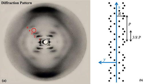
Photograph of the DNA diffraction pattern in its so-called B-configuration
The primary research method involves directing a narrow beam of X-rays at a crystalline sample using an X-ray camera. The resulting photograph displays the pattern of diffracted X-rays passing through the crystal, allowing scientists to visually map its spatial structure, known as the crystal lattice. The various techniques for conducting this method are called X-ray crystallography.


Most viewed


